
Terms to know
AortaThe largest artery in the body. It carries oxygen-rich blood away from the heart to vessels that reach the rest of the body.
CapillariesThe smallest of the body's blood vessels. Oxygen and glucose pass through capillary walls and enter the cells. Waste products such as carbon dioxide pass back from the cells into the blood through capillaries.
Cardiac Valves (Heart Valves)Any of the four heart valves that regulate the flow of blood through the chambers of the heart.
Deoxygenated BloodOxygen-poor blood.HeartThe hollow, muscular organ that maintains the circulation of the blood.
Interventricular SeptumInterventricular septum is the stout wall separating the lower chambers (the ventricles) of the heart from one another.
LungsOne of a pair of organs in the chest that supplies the body with oxygen, and removes carbon dioxide from the body.
MyocardiumThe muscular substance of the heart; the middle of the three layers forming the outer wall of the human heart.
Oxygenated BloodOxygen-rich blood.
Pulmonary ArteryThe pulmonary artery and its branches deliver blood rich in carbon dioxide (and lacking in oxygen) to the capillaries that surround the air sacs.
Pulmonary ArteryThe pulmonary artery and its branches deliver blood rich in carbon dioxide (and lacking in oxygen) to the capillaries that surround the air sacs.
Pulmonary CirculationThe circulation of the blood through the lungs.
Pulmonary VeinsThe veins that return the oxygenated blood from the lungs to the left atrium of the heart.
Pulmonary VeinsThe veins that return the oxygenated blood from the lungs to the left atrium of the heart.
Superior Vena CavaThe large vein that carries blood from the head, neck, arms, and chest to the heart.
Vena CavaA large vein which returns blood from the head, neck and extremities to the heart.
Vena CavaA large vein which returns blood from the head, neck and extremities to the heart.
The blood circulatory system(cardiovascular system) deliversnutrients and oxygen to all cells in the body. It consists of the heartand the blood vessels running through the entire body.
How does the blood circulatory system work?
The blood circulatory system(cardiovascular system) deliversnutrients and oxygen to all cells in the body. It consists of the heartand the blood vessels running through the entire body.
Arteries and Veins
Thearteries carry blood away from the heart; the veins carry it back to the heart. The system of blood vessels resembles a tree: The “trunk,” the main artery (aorta), branches into large arteries, which lead to smaller and smaller vessels.
Thearteries carry blood away from the heart; the veins carry it back to the heart. The system of blood vessels resembles a tree: The “trunk,” the main artery (aorta), branches into large arteries, which lead to smaller and smaller vessels.
The smallest arteries end in a network of tiny vessels, the capillarynetwork.
STRUCTURE OF THE HEART
The human heart is a four-chambered muscular organ, shaped and sized roughly like a man's closed fist with two-thirds of the mass to the left of midline.
The heart is enclosed in a pericardial sac that is lined with the parietal layers of a serous membrane. The visceral layer of the serous membrane forms the epicardium.
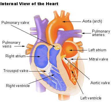
Layers of the Heart Wall
Three layers of tissue form the heart wall. The outer layer of the heart wall is the epicardium, the middle layer is the myocardium, and the inner layer is the endocardium.
Chambers of the Heart
The internal cavity of the heart is divided into four chambers:
- Right atrium
- Right ventricle
- Left atrium
- Left ventricle
The two atria are thin-walled chambers that receive blood from the veins. The two ventricles are thick-walled chambers that forcefully pump blood out of the heart. Differences in thickness of the heart chamber walls are due to variations in the amount of myocardium present, which reflects the amount of force each chamber is required to generate.
The right atrium receives deoxygenated blood from systemic veins; the left atrium receives oxygenated blood from the pulmonary veins.
Valves of the Heart
Pumps need a set of valves to keep the fluid flowing in one direction and the heart is no exception. The heart has two types of valves that keep the blood flowing in the correct direction. The valves between the atria and ventricles are called atrioventricular valves (also called cuspid valves), while those at the bases of the large vessels leaving the ventricles are called semilunar valves.
The right atrioventricular valve is the tricuspid valve. The left atrioventricular valve is the bicuspid, or mitral, valve. The valve between the right ventricle and pulmonary trunk is the pulmonary semilunar valve. The valve between the left ventricle and the aorta is the aortic semilunar valve.
When the ventricles contract, atrioventricular valves close to prevent blood from flowing back into the atria. When the ventricles relax, semilunar valves close to prevent blood from flowing back into the ventricles.
Pathway of Blood through the Heart
While it is convenient to describe the flow of blood through the right side of the heart and then through the left side, it is important to realize that both atria and ventricles contract at the same time. The heart works as two pumps, one on the right and one on the left, working simultaneously. Blood flows from the right atrium to the right ventricle, and then is pumped to the lungs to receive oxygen. From the lungs, the blood flows to the left atrium, then to the left ventricle. From there it is pumped to the systemic circulation.
Blood Supply to the Myocardium
The myocardium of the heart wall is a working muscle that needs a continuous supply of oxygen and nutrients to function efficiently. For this reason, cardiac muscle has an extensive network of blood vessels to bring oxygen to the contracting cells and to remove waste products.
The right and left coronary arteries, branches of the ascending aorta, supply blood to the walls of the myocardium. After blood passes through the capillaries in the myocardium, it enters a system of cardiac (coronary) veins. Most of the cardiac veins drain into the coronary sinus, which opens into the right atrium.
TYPES OF CIRCULATION
1.pulmonary circulation
2.systemic circulation
2.systemic circulation
Pulmonary circulation

Diagram of pulmonary circulation. Oxygen-rich blood is shown in red; oxygen-depleted blood in blue.
Pulmonary circulation is the portion of the cardiovascular system which carries deoxygenated blood away from the heart, to the lungs, and returns oxygenated (oxygen-rich) blood back to the heart.
A separate system known as the bronchial circulation supplies blood to the tissue of the larger airways of the lung.
Route
Pulmonary circulation is the movement of blood from the heart, to the lungs, and back to the heart again.
Deoxygenated blood leaves the heart, goes to the lungs, and then re-enters the heart;
Deoxygenated blood leaves the heart, goes to the lungs, and then re-enters the heart;
Deoxygenated blood leaves through the right ventricle through the pulmonary artery. From the right atrium, the blood is pumped through the tricuspid valve (or right atrioventricular valve), into the right ventricle.
Blood is then pumped from the right ventricle through the pulmonary valve and into the pulmonary trunk of the pulmonary artery.
Arteries
From the right ventricle, blood is pumped through the pulmonary semi-lunar valve into the left and right pulmonary arteries (one for each lung) and travels through the lungs.
Lungs
The pulmonary arteries carry deoxygenated blood to the lungs, wherecarbon dioxide is released and oxygen is picked up during respiration.
Their function is to assist in the carrying of blood to all cells of the body. The pulmonary vein returns oxygenated blood to the left atrium of the heart.
Veins
The oxygenated blood then leaves the lungs
through pulmonary veins, which return it to the left heart, completing the pulmonary cycle.
through pulmonary veins, which return it to the left heart, completing the pulmonary cycle.
This blood then enters the left atrium, which pumps it through the bicuspid valve, also called the mitral or left atrioventricular valve, into the left ventricle.
From the left ventricle the blood passes through the aortic valve to the aorta. The blood is then distributed to the body through the systemic circulation before returning again to the pulmonary circulation.
SYSTEMIC CIRCULATION
Systemic Circuit
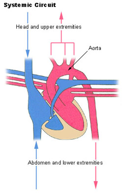
Thesystemic circulation provides the functional blood supply to all body tissue. It carries oxygen and nutrients to the cells and picks up carbon dioxide and waste products. Systemic circulation carries oxygenated blood from the left ventricle, through the arteries, to the capillaries in the tissues of the body. From the tissue capillaries, the deoxygenated blood returns through a system of veins to the right atrium of the heart.
The coronary arteries are the only vessels that branch from the ascending aorta. The brachiocephalic, left common carotid, and left subclavian arteries branch from the aortic arch. Blood supply for the brain is provided by the internal carotid and vertebral arteries. The subclavian arteries provide the blood supply for the upper extremity. The celiac, superior mesenteric, suprarenal, renal, gonadal, and inferior mesenteric arteries branch from the abdominal aorta to supply the abdominal viscera. Lumbar arteries provide blood for the muscles and spinal cord. Branches of the external iliac artery provide the blood supply for the lower extremity. The internal iliac artery supplies the pelvic viscera.
Major Systemic Arteries
All systemic arteries are branches, either directly or indirectly, from the aorta. The aorta ascends from the left ventricle, curves posteriorly and to the left, then descends through the thorax and abdomen. This geography divides the aorta into three portions: ascending aorta, arotic arch, and descending aorta. The descending aorta is further subdivided into the thoracic arota and abdominal aorta.
Major Systemic Veins
After blood delivers oxygen to the tissues and picks up carbon dioxide, it returns to the heart through a system of veins. The capillaries, where the gaseous exchange occurs, merge into venules and these converge to form larger and larger veins until the blood reaches either the superior vena cava or inferior vena cava, which drain into the right atrium.
No comments:
Post a Comment